Contents
As part of the development of MyTwin Lab, we have taken a new step in building the patient’s digital twin: the creation of a 3D anatomical model from a full-body PET scan.
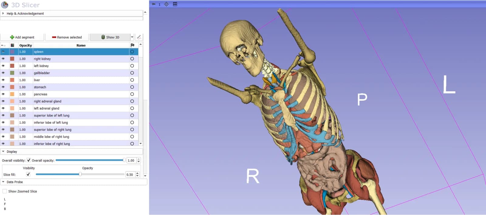
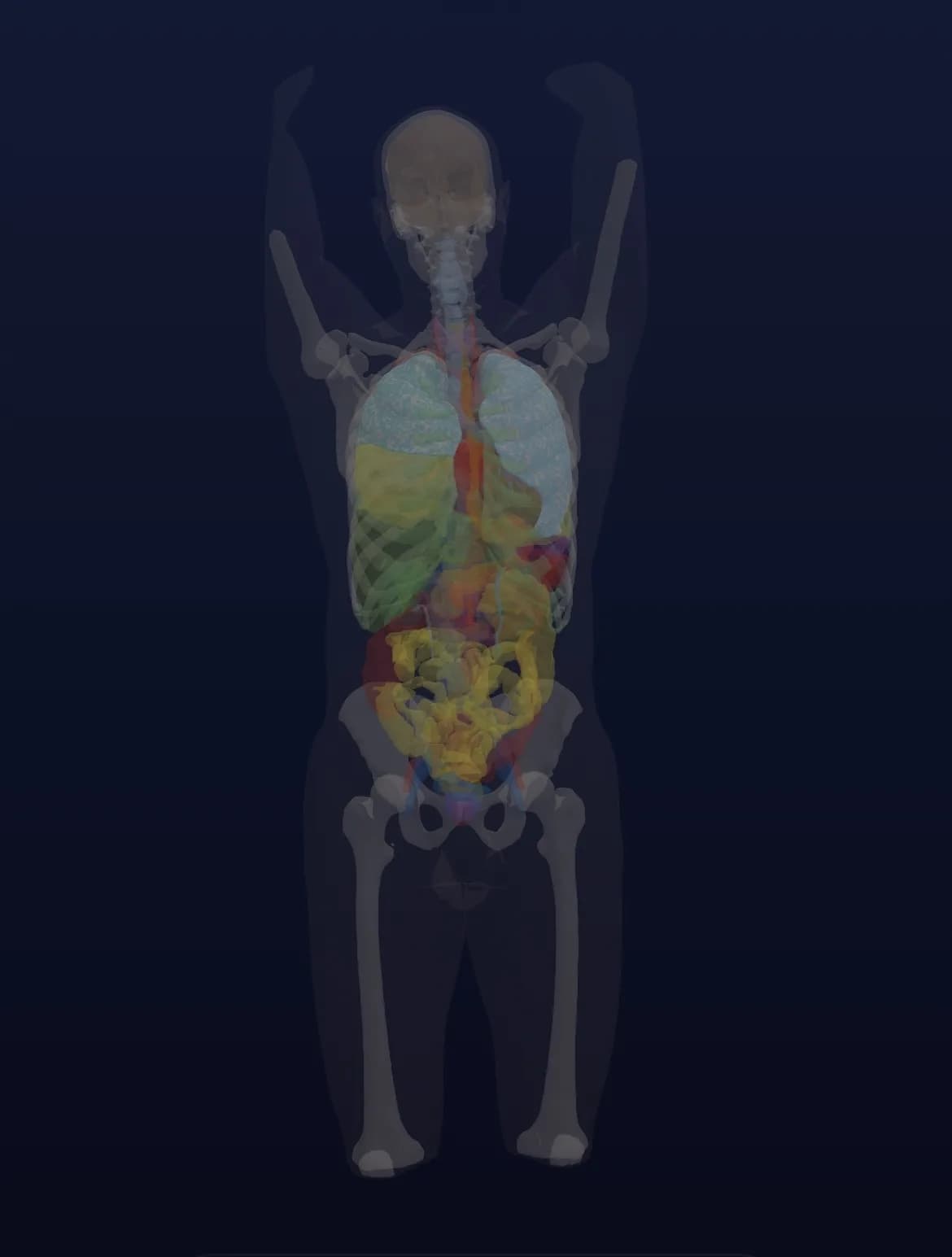
Starting from the medical imaging data, the different anatomical structures were segmented, then reconstructed in three dimensions, to obtain an individualised representation of the patient’s anatomy, especially the thorax and the organs visible on the exam.
 Segmentation
This approach is one of the fundamental building blocks of a health digital twin: starting from a patient’s real data to gradually build a digital representation that is their own. Scientific work on medical digital twins describes exactly this logic: an individualised virtual representation, combined with the patient’s own data and, eventually, with analysis and simulation capabilities.
Seeing the patient’s anatomy differently
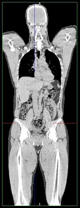
3D reconstruction turns a series of cross-section medical images into a much more intuitive spatial representation of the anatomy.
 A slice
 The 3D model
It can help to better understand the anatomical variations specific to each patient, the relationships between organs, and where certain areas of interest sit in space.
Personalised anatomical modelling is already being studied in several medical fields, notably for therapeutic or surgical planning and for building patient-specific models.
In oncological imaging, PET combined with CT provides complementary functional and anatomical information. 3D reconstruction obviously does not replace the medical reading of a PET scan, but it can add a layer of visualisation and analysis around the patient’s data. What AI can and can’t do with medical images is covered in MyTwin’s guide to AI in medical imaging.
A first step towards a model able to simulate
At MyTwin Lab, though, the goal goes beyond 3D visualisation.
A true digital twin is more than a graphic representation of the body. The scientific literature describes the medical digital twin as an individualised model that can gradually integrate different sources of data, to contribute to monitoring, prediction and the simulation of health scenarios.
Anatomical modelling is therefore a first layer, alongside the outer envelope of the body, captured in a full-body 3D avatar.
Over time, it can be enriched with other information: biological data, medical history, biomarkers, physiological data, imaging, functional data or predictive models.
The challenge is to move, step by step:
- BeforeThe image of the patientA series of cross-section images from the exam.
- This stepTheir 3D modelStructures segmented, then reconstructed in three dimensions.
- To comeA dynamic modelEnriched with other data, able to evolve over time.
Building the most advanced patient digital twin possible
MyTwin Lab’s ambition is to gradually connect these building blocks into an ever more complete and individualised representation of the patient, as described in MyTwin Lab’s research vision.
This representation could, eventually, support different kinds of simulation: how a state of health evolves, the potential response to certain treatments, preparing interventions, comparing scenarios or identifying some risks early.
Current research on medical digital twins explores exactly these applications, notably in personalised medicine, oncology and individual risk prediction. What a patient digital twin is, and what it is for, is explained in MyTwin’s guide to the patient digital twin.
This reconstruction from a PET scan is therefore one more step towards the goal MyTwin Lab pursues: building a digital twin of the human body that is ever more precise, personalised and able, tomorrow, to simulate different possible futures for the same patient.
Frequently asked questions
Does the 3D model replace the reading of the PET scan?
No. The PET scan is read and interpreted by physicians. The 3D reconstruction adds a layer of visualisation and analysis around the patient’s data; it makes no diagnosis.
Can I get a 3D model of my own scans in MyTwin?
Not today. This reconstruction is a MyTwin Lab experiment, and no 3D model of medical images is offered to MyTwin users yet.
Sources
Sources · 3
- 01Katsoulakis E. et al., 2024, “Digital twins for health: a scoping review”, npj Digital Medicine.
- 02Laubenbacher R. et al., 2024, “Digital twins in medicine”, Nature Computational Science.
- 03Shen M. et al., 2024, “The effectiveness of digital twins in promoting precision health across the entire population: a systematic review”, npj Digital Medicine.
This news is provided for information only. The technologies it describes are at research or pilot stage, and none of them replaces advice, diagnosis or treatment from a healthcare professional.